In medical imaging, image registration is the process of transforming different sets of data into one coordinate system, i.e. projecting images into a common reference framework. Clinical data could include magnetic resonance imaging (MRI) scans, computed-tomography (CT), laser surface scans (Surf3D), multiple photographs; also, it may involve data by combining different sensors/cameras, at different time points, depths or even viewpoints. As far as breast cancer management is concerned, this usually involves acquisition of MRI and CT data. Specifically, prone-to-supine breast image registration can be applied into surgical and radiotherapy planning, image-guided interventions, multi-modal cancer diagnosis, staging, and prognosis (response to therapy prediction). However, registration of 3D image data, acquired with the patient in different postures is a challenging problem, due to large deformations induced to the soft breast tissue caused by the change in gravity. To circumvent this, we have integrated biomechanical in silico models with image processing techniques into a registration framework such that it aligns pertinent (MRIs, CTs, Surface) images in a central (reference) configuration by minimizing the target registration error to the image resolution accuracy.
This work was carried out at UCL’s Centre for Medical Image Computing, together with Dr B. Eiben, Dr J. Hipwell and Professor David Hawkes (UCL), and partners from PHILIPS (Germany), Royal Free Hospital (UK) and Champalimaud Foundation (Portugal).
Selected relevant works:
Eiben et al. 2015. Annals of Biomedical Engineering, doi: 10.1007/s10439-015-1496-z
Eiben et al. 2016. SPIE Medical Imaging, doi:10.1117/12.2216728
Eiben et al. 2016. In Lecture Notes in Computer Science, Springer, pp.274-281


https://youtu.be/ljuJozYO2Uc ( cannot be embeded)
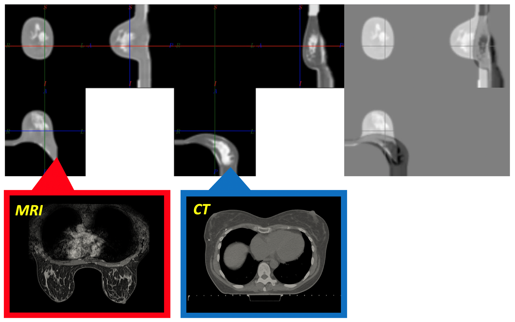
RIGHT: Animation of the registration algorithm with MRI and CT images starting from corresponding breast configurations, which both ultimately converge into a reference ”zero-gravity” configuration to accomplish registering the image data.
https://youtu.be/qHocB2-Kp-A ( cannot be embed into the websites)
Breast conservative surgery combined with radiotherapy has become the treatment of choice for the majority of women presenting with early breast cancer. Due to widespread screening an important percentage of early breast cancers are non-palpable, thus, tumour localization is carried out to “guide” the surgeon during surgery so as to successfully excise and resect the breast lesion. Existing however modalities for pre-operative localization are invasive requiring imaging guidance (e.g., wire-guided, carbon tattooing, radioactive seed localization, etc.). To overcome this, we are developing an innovative technology that is capable fusing augmented reality with in silico and imaging models.
Together with colleagues from the Champalimaud Foundation, INESC (Portugal) and Microsoft, we presented our work in Breast. In this paper we describe the first experimental test with a digital non-invasive method for intra-operative breast cancer localization using AR to guide breast conservative surgery.
Relevant work:
Gouveia et al. 2021. Breast, doi: 10.1016/j.breast.2021.01.004
Vavourakis et al. 2016. PLOS ONE, doi: 10.1371/journal.pone.0159766


